Interview with Dr. Valentina Garibotto
Why did you decide to move into the molecular neuroimaging field?
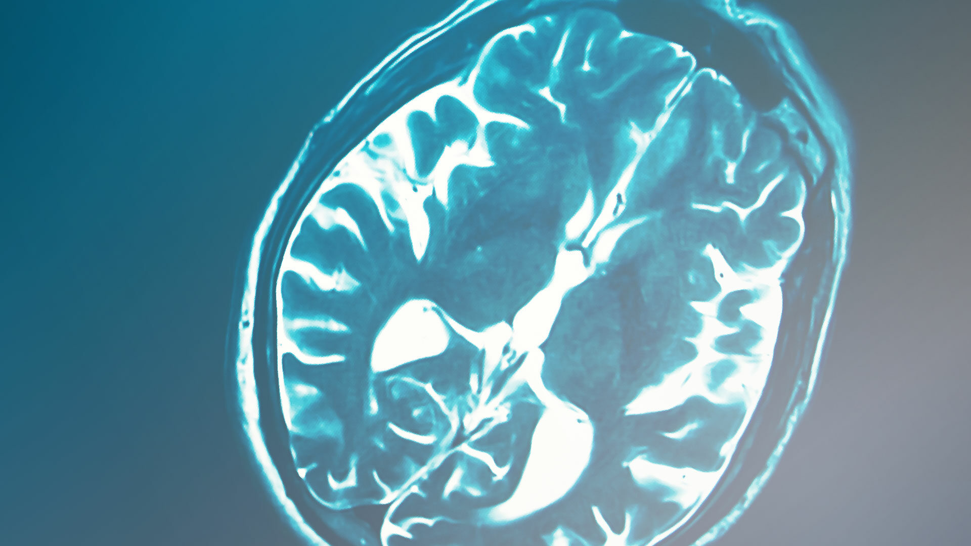
I have always been fascinated by the functioning of our brain and when I discovered that PET imaging could “see” processes and neurotransmittors, I fell in love.
What are the most important findings you discovered so far thanks to your/your groups research activities?
Our main results show that molecular imaging in individuals investigated for cognitive impairment allows to detect the disease earlier and more accurately, to improve the diagnostic process and to identify factors that can modulate the severity of the disease expression. We studied different methods and strategies to evaluate molecular imaging markers, to compare them and to measure them accurately.
Here some references for our main works on the topic:
- Sala et al., 2022
- Giannakopoulos et al., 2021
- Boccardi et al., 2021
- Altomare et al., 2021
- Wolters et al., 2021
- Chétalat et al., 2020
What are the most common used methods/modalities to diagnose Alzheimer’s disease (AD)?
Most common: MRI (Magnetic Resonance Imaging) and CSF (cerebrospinal fluid measurement of amyloid and tau pathologies). Closely followed by PET, most commonly FDG PET but also Amyloid PET.
Can you describe an ideal pathway for AD diagnostic?
The pathway described in this paper is the most recent consensus describing an ideal pathway taking PET into account: Chételat et al., 2020.
Brain PET is not very often used to diagnose AD in Switzerland, what could be the reason for this?
The main limiting factors are:
– the current quite restrictive criteria for reimbursement
– the costs of the procedure
Do you know of other countries where brain PET is a standard diagnostic tool to diagnose AD?
The use is probably comparable across Europe, with local specificities. A list of reimbursement of biomarkers in AD across European countries was summarized here: Frisoni et al., 2017.
What is the advantage of brain PET compared to other diagnostic modalities in AD diagnostic?
FDG PET has higher sensitivity than MRI to assess neurodegeneration in the early neurodegenerative phases (Caminiti et al., 2018), amyloid PET and tau PET are the most accurate diagnostic methods to measure the two hallmarks of AD, also in comparison with other methods to measure amyloid and tau in the cerebrospinal fluid (f.e. Ramusino et al., 2020; Wolters et al., 2020).
How do you foresee AD diagnostic developing in the future?
A combination of imaging and plasma markers in an integrated diagnostic approach (Garibotto et al., 2021): plasma based assessment for screening, PET evaluation in intermediate cases (which I expect to be a large proportion) and in all cases where the topography of brain pathology can be relevant (severity staging, f. e., or atypical presentations) and when a proof of brain pathology for treatment initiation and monitoring is needed.
How do you foresee AD treatment modalities developing in the future?
The approval of aducanumab, despite the debate (Garibotto et al., 2021 ), has shown the importance of PET imaging for selecting populations for treatments and for measuring their biological effects. The data published on more recently developed medication rely even more on the use of PET for the selection of patients to be included in the trial and for the exploration of the biological effects of the drug (Lowe et al., 2021; Klein et al., 2021; Klein et al., 2019; Swanson et al., 2021)
What would you like to accomplish next with your research?
I would like to position our center and group as a reference for the clinical translation of molecular imaging markers in neurodegenerative conditions, for diagnosis and treatment monitoring. These are exciting times for the field and I am convinced we can truly help patients on a daily basis with our methods and tools.
About Dr. Garibotto
Pr. Valentina Garibotto is division chair of nuclear medicine and molecular imaging at the University Hospitals of Geneva and associate professor at the Geneva University. She trained in Italy and Germany and then developed her clinical, research and teaching activity in Geneva. Her project, mainly funded by the Swiss National Research foundation, the Velux Stiftung, the Schmidheiny and Aetas foundation, investigate molecular imaging, namely to guide diagnosis in neurodegenerative conditions, to discover causative and protecting factors and to support the development of novel therapeutic approaches. She has authored more than 190 peer reviewed articles (H index: 32, Web of Science) in the fields of clinical neuroimaging and nuclear medicine. She is Congress Chair Elected of the European Association of Nuclear Medicine, Chair of the Neuroimaging group of the Swiss Society of Nuclear Medicine and actively involved in a number of European initiatives developing and validating molecular imaging in cognitive disturbances.