Interview with Prof. em. Alfred Buck
When did you realize that a dedicated brain PET device is needed?
Already a long time ago. I worked at the University Zurich Hospital PET center as clinician and researcher. I was mostly interested in the brain and PET offered completely new opportunities to explore the brain. At Zurich we were always equipped with the latest technology PET scanners. In a clinical PET center, the scanners are fully booked with patients during the day. Research has to be done in the evenings and weekends. Thirty years ago this was no problem – I could just walk in on weekends and use the scanners and start the cyclotron to produce the needed radionuclides. But due to regulations that became more and more difficult over the years and “weekend research” was almost not possible anymore. Furthermore, the existing whole body PET scanners are expensive and mostly occupied with patients needing whole body scans. Brain clinical and research work could easily be done on more dedicated and cheaper scanners. Such a brain scanner has several important advantages:
a) it could be much cheaper compared to the whole-body scanners
b) The space requirements would be much less. The whole-body scanners need a big room with complex infrastructure. A simple dedicated brain PET scanner, where the patients could be scanned in a sitting position, could be placed in small room next to the big scanners.
c) Such an arrangement would then open many more slots for brain scanning, clinical and for research.
How did PET imaging evolve over time?
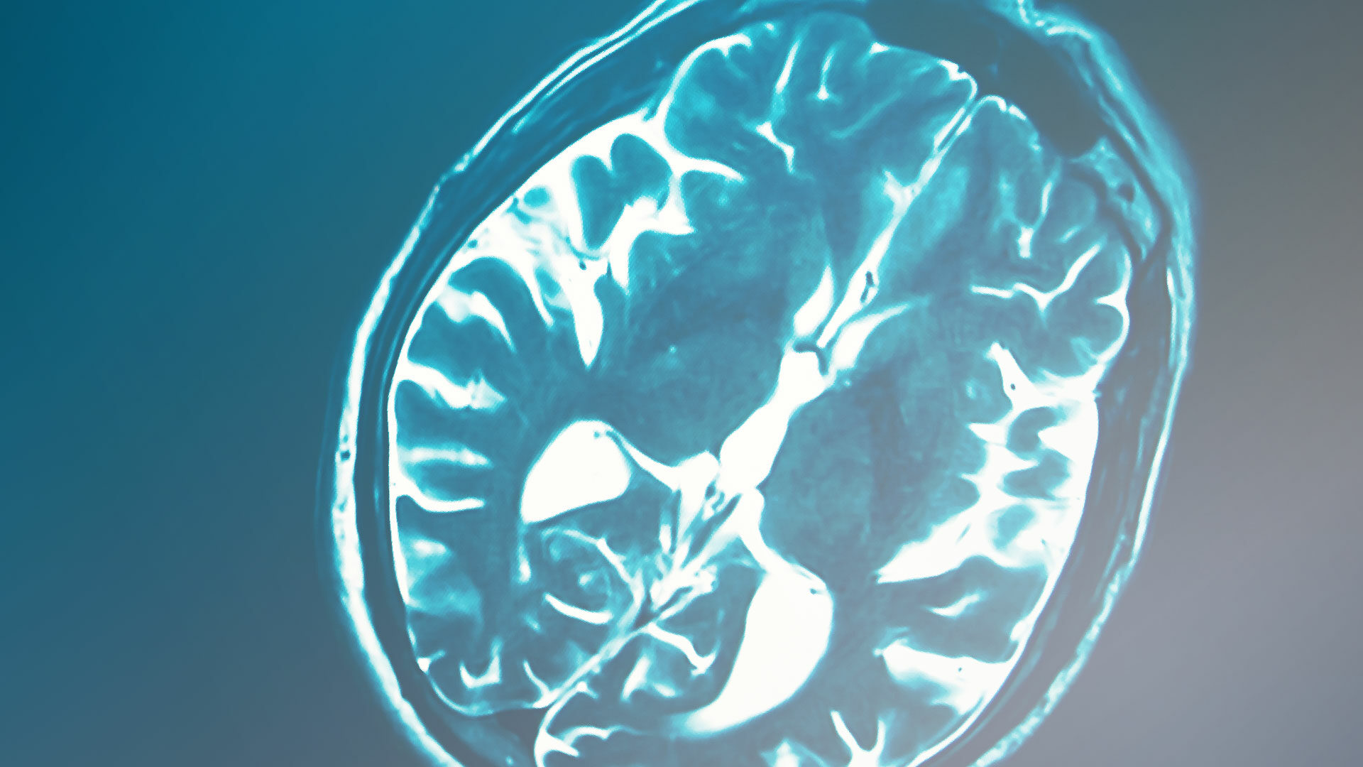
That is an interesting story. Before the year 2000, PET was mainly a research tool. The early PET scanners had a minimal field of view (FOV), like 1 cm, so only a small slice of the body could be imaged. That was fine for brain research, where you get a lot of information in only one slice. The other crucial development was the discovery of Fluorodeoxyglucose (FDG), which could be labelled with F-18. FDG is a glucose analogue, it enters the cell like glucose, gets phosphorylated once but then is not further metabolized and gets trapped in the cell. That makes it ideal for imaging. Even today FDG is the most used PET tracer. Over time the PET scanners got a larger FOV and it became feasible to image the whole body, making the method interesting in cancer diagnosis. A whole-body scan back then took in the order of 1 hour, which was still somewhat impractical. So, the demand for clinical PET scanners remained limited. At around the year 2000 some big manufacturers thought about leaving the PET area because it was not lucrative enough. Then came the game changer. The crucial idea was to combine PET with computerized tomography (CT) in a single machine. This combination allowed to shorten the time for whole body scanning to 20 minutes and less. This was the beginning of an incredible boom in clinical PET imaging. At a later stage, PET was also combined with magnetic resonance imaging (MRI). This combination of different modalities pushed the price of the hardware to millions. Although a valuable tool for whole body imaging, these new machines are somewhat an overkill for brain imaging for several reasons. That is why a dedicated brain scanner like the NeuroLF would be much more economical without a loss in image quality. Such a scanner is much smaller and for brain imaging the hardware combination with a CT or MR is not necessarily needed. For a PET center such a dedicated brain scanner could be an ideal addition to a whole-body scanner.
How do you rate PET imaging in dementia diagnostics compared to other diagnostic dementia modalities?
There are dementias in younger and older people. Younger people have often treatable diseases, like Vitamin deficiencies or thyroid dysfunction. In older people the most prevalent form is Alzheimer’s disease (AD), followed by vascular dementias, Lewy body dementia and fronto-temporal dementia. Therapeutic options for all these are limited or non-existent at present. Some of the dementias show typical pathological changes. In AD one finds pathological deposition of amyloid and tau. Although these play an important role in the progression of AD the exact mechanism is at present a hot topic. Just recently the FDA approved an antibody treatment aimed at removing the amyloid. This treatment (aduhelm) was developed in Zurich and demonstrated promising results in a phase 2 trial. In the larger phase 3 trial the results were less convincing. At present there are further ongoing trials aimed at amyloid and recently also tau.
In this respect it is important to note that both amyloid and tau can be imaged and quantitatively measured using PET. That renders PET an invaluable tool in research and diagnostics of dementias.
The standard diagnostic pathway in dementias is as follows: The first step is a thorough clinical, neurological and neuropsychologial examination to quantify the cognitive decline. These examinations usually yield clear indications regarding the etiology of the dementia, however with limited accuracy. Most important is not to miss treatable causes, such as for instance hydrocephalus. To exclude such causes, MRI imaging is helpful and widely available. Regarding AD there exist treatments which aim at boosting certain neurotransmitter systems and if used properly, these may slow the progression. However, these treatments should only be given if the diagnosis is clear. Here PET is becoming more and more important. A standard procedure today is a brain PET with F-18 FDG, which measures glucose metabolism in the brain and demonstrates a very typical pathologic pattern in AD. Clinical examination combined with FDG PET most often yields a clear diagnosis, which renders further diagnostic procedures superfluous.
How do you see the role of amyloid and tau PET?
Imaging of the amyloid depositions has been possible for decades. For the first time it became possible to visualize and measure the amount of amyloid deposition in the living human brain. It offered new insights into the pathology of Alzheimer- and other neurodegenerative brain diseases. With the advent of therapies aimed at removing or preventing the build-up of amyloid in the brain, amyloid PET became even more important. In the development phase of these therapies, PET allowed to measure the effect of the therapies on the amyloid load. The application of these anti-amyloid therapies will require a diagnostic procedure that demonstrates the presence of amyloid, and for this PET will be an important tool. The pathophysiology of AD is complex and amyloid is certainly only part of the story. Another important player is the deposition of tau, which can also be imaged with PET. Therapies aimed at tau are also in development. It may well be that a combination of amyloid and tau therapies may be more effective than addressing each one separately.
Can you describe a potential future diagnostic pathway in dementias of the elderly?
If treatments aimed at amyloid and/or tau prove effective and become widely available a reasonable diagnostic procedure might be as follows:
After clinical tests people with signs of dementia may get screened with a blood test for amyloid and tau. Such tests are in development and will be cost-effective. If the test is positive, a brain PET will follow to exclude the false positive cases. If also PET is positive, treatment may start.
Amyloid and tau depositions start to build up years before the onset of symptoms. If it could be shown that stopping the build-up of these proteins can prevent AD then the above screening procedure could even be extended to everybody above a certain age.
Where do you see the future of brain PET?
Beyond the future in dementia imaging, PET will continue to play a pivotal role in basically all brain diseases: neurovascular disease, brain tumors, neurodegenerative diseases and epilepsy to mention the most common. Again, there are clear clinical indications to perform a brain PET in these pathologies and there is a huge field of research.
What do you think about NeuroLF?
NeuroLF being a dedicated brain scanner will lower the cost of brain scans. This will be most important if the volume of brain scans increases dramatically as can be expected in dementia diagnostics. I can even imagine a dementia scanning center with multiple brain scanners. Furthermore, due to low spatial requirements it can easily be added to conventional PET-CT or PET-MR whole body scanners and therefore increase the availability of brain scanning slots, for research and clinical applications.
Prof. em. Alfred Buck
Alfred Buck studied mechanical engineering at the ETH in Zurich, then worked as engineer in Canada for 2 years. He then studied medicine at the University of Zurich and got his MD 1987. He joined Nuclear Medicine at the University Hospital Zurich 1987. At Ann Arbor in Michigan he did a 2 year fellow-ship in NeuroPET under the auspices of David Kuhl, one of the leading researchers in the field of Alzheimer’s disease. After 2 more years as resident in Neurology, he finished his residency in Nuclear Medicine and became Head of NeuroPET at the University Hospital. He got several grants from the Swiss National Science Foundation and authored many peer reviewed papers. Besides clinical and research work he invented an automatic injector for radioactive substances, the patent of which is licensed to the injector Division of Bayer. He also co-founded the company Swisstrace which sells blood sampling devices for research using PET.